 |
 |
 |
 |
 |
 |
| BULKY UTERUS |
| [Enlarged Uterus] |
Diffuse uterine enlargement is a common clinical finding. Because this abnormality can represent a physiologic manifestation, benign tumor, or malignancy, the diagnostic dilemma of a diffusely enlarged uterus can be challenging. Clinical findings can provide valuable information in regard to physiologic effects, pregnancy-related changes, and hormonal causes. Cytologic examination is essential for identification of cervical and endometrial malignancies. However, since preoperative histologic examination of myometrial lesions is not possible, preoperative distinction between benign and malignant conditions is frequently difficult. Imaging thus plays an important role in evaluation of myometrial lesions. In particular, magnetic resonance (MR) imaging allows specific diagnosis of several different lesions. Signal voids and prominent vessels at MR imaging are characteristic of vascular lesions. Adenomyosis and leiomyomas can be distinguished from other lesions with MR imaging, although a variety of unusual manifestations can be seen. MR imaging findings that allow distinction between leiomyoma and leiomyosarcoma have yet to be clearly established; however, invasion, hemorrhagic necrosis, or rapid growth is suggestive of malignancy. Endometrial stromal sarcoma tends to have distinct MR imaging features that allow differentiation from benign lesions. |
|
Introduction:
Various conditions manifest as diffuse uterine enlargement, which represents one of the most common gynecologic findings. In adult women, the normal uterine corpus is approximately 5.0 cm in height (length), 5.0 cm in width, and 2.5 cm in anteroposterior thickness .This article discusses the uterus that is obviously larger than this size. Potential causes include physiologic changes, benign conditions, and malignant tumors. The myometrium is commonly involved, although the endometrium can also be involved. Imaging has an especially important role in evaluation of myometrial lesions, as histologic samples are not available for such lesions. |
|
Increased thickness of the endometrium and myometrium. The signal intensity of the outer myometrium is increased |
Hormonal Causes:
An enlarged uterus is frequently encountered in the presence of hormonal abnormalities. The most important hormonal factors resulting in uterine enlargement are increased serum levels of estrogen, progesterone, and gonadotropin. Uterine exposure to these hormones can be endogenous (eg, hormone-producing tumors) or exogenous.
The clinical presentation and history are important for identification of these conditions. Examples are a history of drug administration and abnormal or prolonged bleeding inconsistent with patient age or menstrual cycle stage. Nonetheless, imaging findings can also be helpful. In conditions resulting from hormonal effects, an enlarged uterus has normal zonal anatomy, although the thickness or signal intensity of the endometrium and myometrium is abnormally increased.
Exogenous Hormones
Estroprogesterone.— Estroprogesterone (oral contraceptive pill) is used for both oral contraception and treatment of dysmenorrhea. Estroprogesterone administration can result in a myometrium that may appear swollen and globular with high signal intensity, whereas the endometrium can become atrophic due to smooth muscle hypertrophy, sinusoidal dilatation, and edema Myometrial swelling and endometrial atrophy are more pronounced in women taking higher concentrations of the hormone
Vascular Lesions:
A bulky uterus may be encountered secondary to abnormal vascular lesions. Prominent gonadal, parametrial, and myometrial vasculature, with or without arteriovenous shunting, characterizes such lesions. Uterine enlargement can result from prominently engorged vessels or myometrial edema . Although angiography is the standard of reference for diagnosing AVMs and other vascular abnormalities, MR imaging and MR angiography are emerging as effective modalities for noninvasive evaluation of such conditions.
Adenomyosis:
The term adenomyosis refers to benign invasion of the endometrium into the myometrium with reactive overgrowth of the musculature. US illustrates the lesion as decreased echogenicity or heterogeneity of the myometrial region with small embedded cysts . MR imaging demonstrates diffuse or focal widening of the junctional zone, with a width of greater than 12 mm considered to be highly associated with adenomyosis.
Bright spots are observed on T1- or T2-weighted images in lesions of adenomyosis . The bright foci correspond to heterotopic endometrial tissue, cystic dilatation of endometrial glands, or hemorrhagic foci. At US, such areas appear as small myometrial cysts within myometrial regions of decreased echogenicity or heterogeneity . |
 |
|
 |
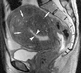
Adenomyosis. Sagittal T2-weighted image shows indistinct zonal anatomy. Widening of the junctional zone is clearly seen in the region around the distorted endometrium (arrowheads). The myometrium has decreased signal intensity with tiny spots of high signal intensity (arrows). |
|
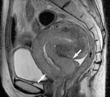
Focal adenomyosis in a 49-year-old woman. Sagittal T2-weighted image shows a heterogeneous area of high signal intensity (arrowheads) within the myometrium that protrudes into the uterine cavity. The interface between the lesion and the myometrium is indistinct. Fine hyperintense striations (arrows) extend into the myometrium; this appearance is an extreme example of pseudowidening of the endometrium. |
|
Neoplasms:
Neoplasms are probably the most common and, at the same time, the most important category of lesions in evaluation of an enlarged uterus. Diffuse neoplastic uterine enlargement typically exhibits two patterns: multiple nodules and extensive replacement of the myometrium by tumor. Mesenchymal or myometrial masses are difficult to evaluate with histologic examination, whereas epithelial tumors are easily evaluated with such studies.
Multiple Nodules
Neoplasms that typically manifest as multiple nodules include leiomyoma, leiomyosarcoma, and endometrial stromal sarcoma. MR imaging findings of uterine leiomyomas are well established . In contrast, reports concerning other mesenchymal tumors are very limited, which thus hinders effective evaluation of an enlarged uterus. |
 |
Leiomyoma:— Leiomyomas are by far the most common uterine tumor, frequently manifesting as diffuse uterine enlargement with multiple nodules. Consistent with their benign nature, leiomyomas exhibit a "pushing" border and a rounded appearance. Leiomyomas typically display distinct low signal intensity with a speckled appearance on T2-weighted images . Although leiomyomas can demonstrate variable appearances due to the presence of edema and hyaline, cystic, and red degeneration, knowledge of the MR imaging characteristics of such variations can aid in differentiating leiomyomas from other mesenchymal tumors.
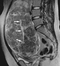
Multiple leiomyomas in a 44-year-old woman. Sagittal T2-weighted image shows a diffusely enlarged uterus with multiple leiomyomas. Each leiomyoma has clear margins and distinct low signal intensity. |
|
Intravenous Leiomyomatosis:— Intravenous leiomyomatosis is a rare condition characterized by leiomyomas remarkable for growth of smooth muscle cells into venous vasculature, although they are otherwise unremarkable. Convoluted, wormlike masses growing within veins, often extending into the broad ligament, other pelvic veins, the inferior vena cava, or even the heart , are the hallmark features of intravenous leiomyomatosis. |
 |
Diffuse Leiomyomatosis:— Diffuse leiomyomatosis is defined as the presence of innumerable small leiomyomas that produce symmetrical enlargement of the uterus, replacing most of the uterine parenchyma . MR imaging demonstrates innumerable nodules that blend with one another and replace the uterine parenchyma to near completion . Such nodules display low to intermediate signal intensity on T2-weighted images. All reported cases necessitate hysterectomy regardless of the patient’s reproductive age, as hormonal treatments have not proved helpful. Nonetheless, uterine artery embolization might be an effective alternative treatment for this benign condition. Definitive diagnosis of this condition with MR imaging might allow consideration of uterine artery embolization as a treatment option for women of reproductive age.
Diffuse leiomyomatosis in a 31-year-old woman. Sagittal T2-weighted image shows a prominently enlarged uterus with innumerable leiomyomas that appear to blend with one another. The endometrium (arrows) is markedly elongated and distorted by multiple submucosal nodules. |
|
 |
Leiomyosarcoma:— Leiomyosarcomas are very rare tumors that are often initially misdiagnosed as leiomyomas. Rapid growth and extensive metastasis are frequently encountered with leiomyosarcomas. As the disease progresses, which can occur with extreme rapidity after an initial manifestation, leiomyosarcomas appear as either a prominently enlarged uterus with multiple sarcomatous nodules or extensive invasion Reported MR imaging findings are variable and include a lobulated mass of high signal intensity on T2-weighted images, a sharply marginated mass of low signal intensity that closely resembles a leiomyoma, or a mass with focally infiltrative margins . Signal intensity is not a reliable indicator of malignancy. Detection of scattered foci of hemorrhage or necrosis can serve as a clue for diagnosis of leiomyosarcoma, as such findings can reflect coagulative necrosis, which should raise suspicion for leiomyosarcoma. At MR imaging, such necrotic areas are seen as areas of slightly high signal intensity on T1-weighted images and heterogeneous areas on T2-weighted images. |
|
Leiomyosarcoma in a 44-year-old woman. (a) Sagittal T2-weighted image shows a tumor (M) with slightly high signal intensity and irregular margins. The tumor protrudes from the posterior myometrium into the endometrial cavity (arrows). Small leiomyomas (m) with clear margins are present in the anterior wall. (b) Sagittal T2-weighted image, obtained 3 months later after the patient experienced rapidly progressing abdominal fullness, shows an irregularly shaped uterus that has clearly increased in size. The tumor occupies the endometrial cavity (arrows). The nodules (m) in the anterior wall also demonstrate remarkable increase in size.
Diffuse Myometrial Involvement:
Diffuse myometrial involvement is usually observed with malignant tumors. Although most lesions in this category diffusely involve the uterus, occasionally nodular involvement can be observed with these lesions.
Hematologic Malignancies:— The uterine cervix and corpus are rarely the primary site for leukemia or lymphoma. When uterine lymphoma does occur, the most common manifestation is diffuse symmetrical uterine enlargement with relatively high signal intensity on T2-weighted images and epithelial preservation . As malignant uterine lymphoma has a fair prognosis if properly treated, accurate evaluation and diagnosis of the disease are essential . Since it is sometimes difficult to distinguish malignant lymphoma from other uterine malignancies with routine biopsy samples, imaging findings can be helpful in making the appropriate diagnosis. |
 |
Malignant lymphoma in a 48-year-old woman with multiple swollen lymph nodes in the paraaortic and supraclavicular regions. (a) Sagittal T1-weighted image shows extensive uniform enlargement of the uterus (arrowheads), which has homogeneous low signal intensity. (b) Sagittal T2-weighted image shows diffuse symmetrical enlargement of the uterus, especially of the cervix (arrows). The lack of signal in the cervical stroma is obvious. The myometrium also lacks its zonal appearance and has low signal intensity with an irregular contour. The cause of this signal intensity is unknown. (c) Sagittal contrast-enhanced T1-weighted image shows heterogeneous enhancement of the uterus. The diagnosis of malignant lymphoma was established by means of biopsy of a lymph node. The uterine lesion significantly decreased in size with chemotherapy and was considered to represent uterine involvement by lymphoma. |
|
 |
Endometrial Stromal Sarcoma:— Endometrial stromal sarcoma originates from the endometrium but invariably exhibits extensive myometrial involvement that is either sharply demarcated or diffusely infiltrative . This entity is subdivided into low-grade stromal sarcoma and high-grade stromal sarcoma. Low-grade stromal sarcoma can be encountered even in the second decade of life. Moreover, the diagnosis is difficult to establish because, in general, the tumor cells are not sufficiently atypical to be accurately distinguished from benign endometrial stromal cells. In contrast, preoperative diagnosis of high-grade stromal sarcoma is readily established by microscopic examination of curettage material. An important diagnostic clue for low-grade stromal sar-coma is identification of bands of low signal intensity within the area of myometrial invasion, which is either sharply demarcated or diffusely infiltrative on T2-weighted images .. Extension along the vessels, fallopian tubes, or ligaments is another characteristic of the tumor. Needless to say, imaging studies play an important diagnostic role in endometrial stromal sarcoma, especially in the case of low-grade stromal sarcoma.
Low-grade endometrial stromal sarcoma in a 21-year-old woman. Sagittal T2-weighted image shows a huge tumor that replaces the endometrial cavity (arrowheads) and infiltrates the myometrium. Bands of low signal intensity (arrows) are seen in the infiltrated myometrium; these bands corresponded to preserved bundles of myometrium at histologic examination. |
|
Other Uterine Malignancies:— Other malignancies may extensively involve the uterus in advanced stages, including cervical and endometrial cancers as well as mixed müllerian tumors. A diagnosis can be established from histologic samples obtained prior to imaging studies. Small cell carcinomas, which can arise from either the cervix or endometrium, are known for their propensity to spread rapidly and systemically and thus bear a poor prognosis. MR images demonstrate uniform uterine enlargement, which mimics the appearance of malignant lymphoma. Myometrial signal intensity is diffusely high on T2-weighted images
Uterine Metastatic Disease:— Metastatic disease to the uterus is rare; if present, it indicates a malignancy in an advanced stage . The most common primary sites are the breast and stomach. On MR images, the uterus appears enlarged. T2-weighted images exhibit abnormally high or low signal intensity throughout the myometrium Differentiating metastatic disease from hematologic diseases, such as lymphoma and leukemia, becomes difficult if the lesions exhibit high signal intensity. In contrast, when the lesions exhibit low signal intensity, a misdiagnosis such as adenomyosis can result.
TREATMENT:
Saravana hospital homoeopathic treatment gives excellent cure for Bulky uterus.. please see the cured reports. |
|
 |
 |
 |
 |
|
 |
2010 ® Saravana Hospital. All rights reserved . |
|

